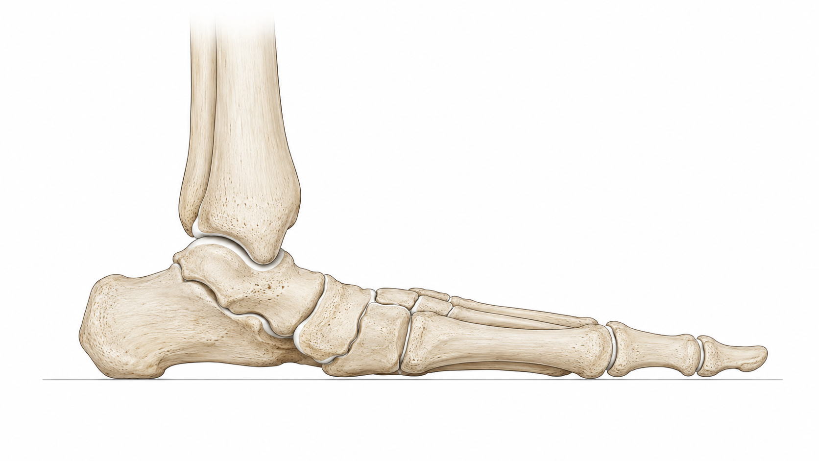
Outcomes Flexible flatfoot
A flexible flatfoot remains mobile and the arch partially or completely reforms when the foot is not bearing weight or when the patient rises onto their toes. Many flexible flat feet are painless and require no treatment.
When symptoms are present, initial management generally involves supportive footwear, foot orthoses, strengthening, activity modification or bracing. These measures may improve comfort and function but do not permanently alter the underlying structure.
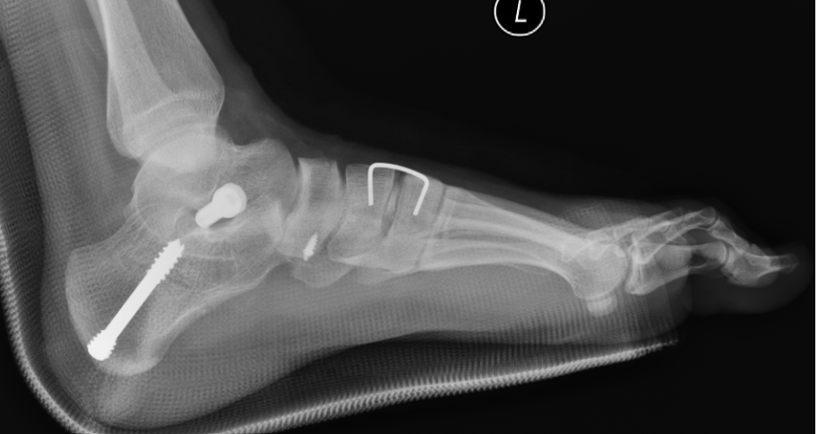
For selected symptomatic flexible deformities, a minimally invasive procedure such as subtalar arthroereisis may be considered. HyProCure® is a small titanium implant placed within the sinus tarsi—the natural space between the talus and calcaneus. It is intended to limit excessive talotarsal displacement while preserving joint movement. Additional procedures, such as gastrocnemius recession, may occasionally be required.
Published HyProCure studies have generally reported improvements in pain and function. One prospective series reported an approximate 37% reduction in pain and improvement in overall Maryland Foot Scores from 69.5 to 89.2 at one year (higher score = better outcome). A separate study of extra-osseous talotarsal stabilisation reported a reduction in average pain from 5.5/10 to 2.2/10, with overall patient satisfaction of 84%.
Sinus tarsi pain is the most frequent complication and may require repositioning or removal of the implant. Published removal rates vary considerably according to the implant, patient group and associated procedures. HyProCure-specific studies have reported removal rates of approximately 1- 4%. The procedure is therefore appropriate only for carefully selected flexible and reducible deformities.
HyProCure prospective outcome study
Extra-osseous talotarsal stabilisation outcomes
Manufacturer’s website: https://gramedica.com/
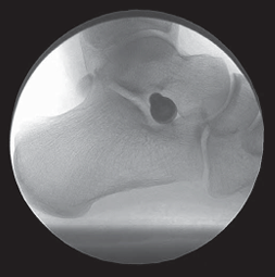
What is a Pes Planus - Flat Foot?
Pes planus, commonly known as flat feet, is a complex three-dimensional deformity involving changes in the alignment of the entire foot. The medial longitudinal arch becomes lower, the heel may tilt outwards, and the forefoot may move away from the body. This can also make the midfoot appear more prominent along its inner border.
Many people have flexible flat feet throughout their lives without pain or functional limitation. A flat foot does not require treatment simply because the arch appears low. Treatment may be required if a flatfoot becomes progressive and increasingly symptomatic.
A more complex or progressive flatfoot deformity can involve multiple joints, bones, tendons and ligaments. The condition may remain flexible or gradually become rigid and arthritic. In adults, progressive collapse may be associated with dysfunction of the posterior tibial tendon and supporting ligaments, although it is now understood to be more complex than a tendon disorder alone.
Symptoms may include:
Pain or swelling along the inside of the foot or ankle
Pain around the outside of the ankle
Fatigue or aching with prolonged standing and walking
Difficulty performing a single-leg heel raise
Progressive loss of arch height
Difficulty fitting footwear or orthoses
Reduced walking or exercise tolerance
Stiffness associated with more advanced or arthritic deformity
Outcomes Complex, severe or rigid flatfoot
A complex flatfoot involves more than a simple reduction in arch height. It may include outward angulation of the heel, abduction of the forefoot, instability or collapse through the midfoot, tendon and ligament insufficiency, joint subluxation and arthritis.
As the condition progresses, the deformity may become increasingly rigid and may no longer correct when the foot is unloaded or when the patient rises onto their toes. These deformities are not generally suitable for correction with a stand-alone arthroereisis implant.
Surgical reconstruction is tailored to the individual pattern of collapse and may involve several procedures, which may involve cutting and repositioning bones, fusing joints, and soft tissue balancing.
Flexible deformities can often be reconstructed using joint-preserving osteotomies and soft-tissue procedures. Severe, rigid or arthritic deformities are more likely to require one or more joint fusions.
Clinical studies generally report meaningful improvements in pain, function and quality of life following appropriately selected flatfoot reconstruction. One study reported improvement in pain, function and health-related quality. However, reconstruction involves a substantially longer recovery than arthroereisis and commonly requires six to eight weeks of non-weight-bearing.
Outcomes depend on the severity of the deformity, the joints involved and the combination of procedures required. The aim is to produce a more stable, better-aligned and less painful foot rather than a visually “perfect” arch.
Adult flatfoot reconstruction outcomes
Adult-acquired flatfoot review
What should I expect after surgery?
Recovery differs considerably between a limited HyProCure® procedure and complex flatfoot reconstruction.
HyProCure® and flexible flatfoot correction
An isolated HyProCure procedure generally takes approximately 20–40 minutes and is commonly performed as day surgery. A small incision is made over the sinus tarsi, and the implant is positioned without cutting or fusing bone.
Patients are generally permitted protected or partial weight-bearing in a surgical sandal or walking boot. This is not equivalent to normal, unrestricted walking, and crutches may be required initially.
Recovery may be longer when HyProCure is combined with gastrocnemius recession or another procedure. Temporary sinus tarsi discomfort is common during early recovery. Persistent pain may occasionally require implant repositioning or removal.
Complex flatfoot reconstruction
Complex reconstruction may involve several osteotomies, tendon or ligament procedures and joint fusions. Surgery may take approximately two to three hours and generally requires a considerably longer recovery.
Most patients require strict non-weight-bearing in a splint, cast or protective boot. Crutches, a knee scooter or another mobility aid will usually be required.
Recovery may be longer when several joints are fused or multiple areas of the foot are reconstructed. Swelling can persist for six to twelve months, and maximum improvement may not be apparent for at least a year.
Return to driving, employment, footwear and exercise should be guided by the procedures performed, clinical review and radiographic healing rather than time alone.